-
Zusatztext
-
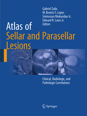
This book presents, in a stepwise and interactive fashion, approximately 75 cases that reflect the wide spectrum of pathology encountered in this region. Each case description commences with a concise clinical scenario. High-quality radiologic, laboratory, and histopathologic images depicting the differentiating features of the lesion subtype in question are then presented, and key operative and clinical management pearls are briefly reviewed. The interdisciplinary nature of this easy-to-use color atlas and textbook reflects the fact that the management of patients with sellar and parasellar lesions is itself often interdisciplinary. The format is unique in that no similar interdisciplinary book is available on lesions of this region of the brain. Atlas of Sellar and Parasellar Lesions: Clinical, Imaging, and Pathologic Correlations is of great value for practitioners and trainees in a range of medical specialties, including radiology, neurology, endocriniology, pathology, oncology, radiation oncology, and neurosurgery.
-
-
Kurztext
-
This book presents, in a stepwise and interactive fashion, approximately 75 clinical-pathological entities that reflect the wide spectrum of pathology encountered in the sellar and parasellar region and have been gathered over a period of decades by the authors. Multiple examples of high-quality radiologic and histopathologic images depicting the differentiating features of the varying lesions are presented, and key operative and clinical management pearls are briefly reviewed in a concise, bullet point format. The interdisciplinary nature of this easy-to-use color atlas and textbook reflects the fact that the management of patients with sellar and parasellar lesions is itself often interdisciplinary,making this atlas a useful resource for any practitioner or trainee who cares for patients with pituitary disease and anterior skull base pathology. The format is unique in that no similar interdisciplinary book is dedicated specifically to the comprehensive catalogue of lesions in this intricate region of the brain and skull base. Atlas of Sellar and Parasellar Lesions: Clinical, Imaging, and Pathologic Correlations is of great value for practitioners and trainees in a range of medical specialties, including radiology, neurology, endocriniology, pathology, oncology, radiation oncology, and neurosurgery.
-
-
Autorenportrait
- Gabriel Zada, MD, MS Department of Neurological Surgery Keck School of Medicine University of Southern California 1975 Zonal Avenue Los Angeles, CA 90033, USAgzada@usc.edu M. Beatriz S. Lopes, MD, PhD Department of Pathology (Neuropathology)University of Virginia School of Medicine1215 Lee Street, Room 3060-HEPCharlottesville, VA 22908, USAmsl2e@virginia.edu Srinivasan Mukundan, Jr, PhD, MDDepartment of RadiologyBrigham and Women's Hospital75 Francis StreetBoston, MA 02115 USA smukundan@partners.org Edward Laws, Jr, MD Department of Neurosurgery Brigham and Women's Hospital Harvard Medical School 15 Francis Street Boston, MA 02115, USA elaws@partners.org
Detailansicht
Atlas of Sellar and Parasellar Lesions
Clinical, Radiologic, and Pathologic Correlations
ISBN/EAN: 9783319342719
Umbreit-Nr.: 2874070
Sprache:
Englisch
Umfang: xviii, 546 S., 242 s/w Illustr., 84 farbige Illust
Format in cm:
Einband:
kartoniertes Buch
Erschienen am 23.08.2016
Auflage: 1/2016


