-
Zusatztext
-
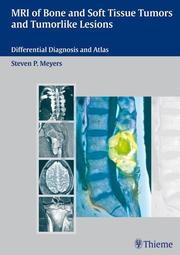
Practical. In-depth. Invaluable. A guide to the diagnosis of tumors and tumorlike lesions of bone and soft tissue using MRI. This unique encyclopedic guide takes the same approach you apply in clinical practice. It features fully illustrated differential diagnosis tables organized according to MRI findings and the locations of tumors. An in-depth reference section provides information on each lesion. In addition, almost 3000 high quality images make this practical text an invaluable tool in the diagnosis of common and rare tumors and other disorders of the musculoskeletal system. Features: 20 differential diagnosis tables based on anatomic locations of lesions rather than disease Fully illustrated reference chapters containing concise, detailed information for each lesion - from relative frequency and age ranges to MRI findings, treatment, and prognosis Over 2900 state-of-the-art illustrations covering the wide range of imaging features for various lesions An exceptional level of detail, helping you to differentiate between diseases and conditions that have similar appearances Extensive cross-referencing to further up-to-the minute resources This is the definitive guide to MRI of musculoskeletal tumors. Whether you need a practical guide for day-to-day use or a comprehensive preparation tool for board examinations - keep this text close to the workstation.
-
-
Kurztext
-
Practical. In-depth. Invaluable. A guide to the diagnosis of tumors and tumorlike lesions of bone and soft tissue using MRI. This unique encyclopedic guide takes the same approach you apply in clinical practice. It features fully illustrated differential diagnosis tables organized according to MRI findings and the locations of tumors. An in-depth referencesection provides information on each lesion. In addition, almost 3000 highquality images make this practical text an invaluable tool in the diagnosis of common and rare tumors and other disorders of the musculoskeletal system. Features: 20 differential diagnosis tables based on anatomic locations of lesions rather than disease Fully illustrated reference chapters containing concise, detailed information for each lesionfrom relative frequency and age ranges to MRI findings, treatment, and prognosis Over 2900 stateoftheart illustrations covering the wide range of imaging features for various lesions An exceptional level of detail, helping you to differentiate between diseases and conditions that have similar appearances Extensive crossreferencing to further uptothe minute resources This is the definitive guide to MRI of musculoskeletal tumors. Whether you need a practical guide for day-to-day use or a comprehensive preparation tool for board examinations-keep this text close to the workstation. Steven P. Meyers, MD, PhD is Professor of Radiology, Imaging Sciences, and Neurosurgery and full-time faculty member at the University of Rochester Medical Center, Rochester, NY. He is also the Director of the AdvancedFellowship in Magnetic Resonance Imaging at the University of Rochester School of Medicine and Dentistry.
-
Detailansicht
MR Imaging of Musculoskeletal Tumors and Tumor-like Lesions
Differential Diagnosis and Atlas
ISBN/EAN: 9783131354211
Umbreit-Nr.: 1144133
Sprache:
Englisch
Umfang: 816 S., 2500 Illustr.
Format in cm:
Einband:
gebundenes Buch
Erschienen am 21.11.2007
Auflage: 1/2007


